Follow the light in the discovery of the wonders of nano
 Ellipsometry is a versatile, non-destructive technique that works in real time in any transparent medium, based on the fact that when light is reflected from a surface of a material its polarization state undergoes some change depending on its own characteristics.
The word ellipsometry was first introduced in a scientific publication by Alexander Rother in 1945, and is based on the fact that the polarization of the light is generally elliptical. In the last years of the XIX century, Paul Drude derived the mathematical equations that allowed the calculation of the optical constants. In the 60’s, due to the availability of computers for numeric processing, ellipsometry suffered a renaissance. Besides measuring the thickness of a film of few nanometers or its optical constants, today, ellipsometry finds applications in different areas like semiconductor and data storage solutions, biosensors, optical coatings, microelectronics, surface chemistry.
") The one recently acquired by INL is a spectroscopic imaging ellipsometer. With spectroscopy it is possible to know the optical constants as a function of the wavelength of light, enlightening to greater detail the secrets of complex materials. The imaging feature enables, for example, analyzing biochips without any labeling of the probes, an advantage when compared with techniques based on fluorescence or radioactivity. It is thus possible to examine the surface structure of, for instance DNA, on substrates like glass, silicon or gold with high spatial resolution and also in real-time to measure adsorption reactions by using diverse solid-liquid cells for in vitro conditions.
The one recently acquired by INL is a spectroscopic imaging ellipsometer. With spectroscopy it is possible to know the optical constants as a function of the wavelength of light, enlightening to greater detail the secrets of complex materials. The imaging feature enables, for example, analyzing biochips without any labeling of the probes, an advantage when compared with techniques based on fluorescence or radioactivity. It is thus possible to examine the surface structure of, for instance DNA, on substrates like glass, silicon or gold with high spatial resolution and also in real-time to measure adsorption reactions by using diverse solid-liquid cells for in vitro conditions.
Follow the light in the discovery of the wonders of nano and be amazed with the beauty!
The photos show INL Engineer, Adelaide Carvalho, the author of this post, monitoring ellipsometry tests. The spectroscopic imaging ellipsometer, recently acquired by INL. Testing the equipment has been her main concern for the last couple of months.
Observing biological complexes at the single molecule level with optical microscopy at room temperature
The contribution of optical microscopy to life sciences can hardly be underestimated. Ever since the popularization of the optical microscope among biologists in the 17th century, mainly fueled by advances made by Anton van Leeuwenhook leading to the observation of single cell organisms, a long journey of technological development started to improve optical resolution over the complete frequency spectrum of light. Nowadays optical microscopes have achieved a lateral and longitudinal resolution around 200 and 600nm respectively. Unfortunately, further improvements are hampered by the diffraction of light waves.
Since the beginning of the 90s several new techniques have been introduced to circumvent the diffraction limitation for far-field optical image formation by fluorescence, light emitted from samples after absorption of excitation light. One of these techniques is based on the detection of single nano-sized fluorescence sources. Recently, a team of researchers under supervision of Steven Chu, Nobel laureate and current United States Secretary of Energy, have improved distance measurements between two molecular fluorescence centers from the 5-20nm range down to 0.77nm thereby bringing optical localization with visible light in the angstrom regime.
This major breakthrough was realized by a thorough analysis of the noise sources that influence the optical localization measurements. Two closed feedback loops were implemented to perform dual color image registration in order to reduce systematic effects such as drift and vibration of the instrumentation. Further, it was postulated that previous attempts to improve resolution were hampered by lack of characterization of the interpixel photoresponse non-uniformity of charge-coupled device (CCD) cameras used in these studies.
As a demonstration of future applications for this new measurement concept, a structural analysis of epithelial cadherin dimers, complexes that mediate cell adhesion but whose molecular functionality remains elusive, was demonstrated. Hereby, it was demonstrated that biological structures can be analyzed with 1-2nm resolution at room temperatures and in physiological buffer conditions. This new technique is likely to provide new insights in biological complexes that cannot be studied with nuclear magnetic resonance (NMR) and electron paramagnetic resonance (EPR) under these conditions.
Pieter De Beule
18/10/10

Pieter de Beule with confocal microscope add-on he is building with his group.
INL is interested in new high resolution imaging instrumentation and started research collaboration in 2009 with the Max Planck Institute for Biophysical Chemistry. In this project, Pieter De Beule, an INL postdoctoral researcher, works on the design and implementation of a new microscope for optical sectioning of live cells under the supervision of Thomas Jovin. This microscope will help scientists to further develop the understanding of basic cell biology, especially by capturing events occurring on fast timescales such as signal transduction.
Second INL Annual Meeting in April
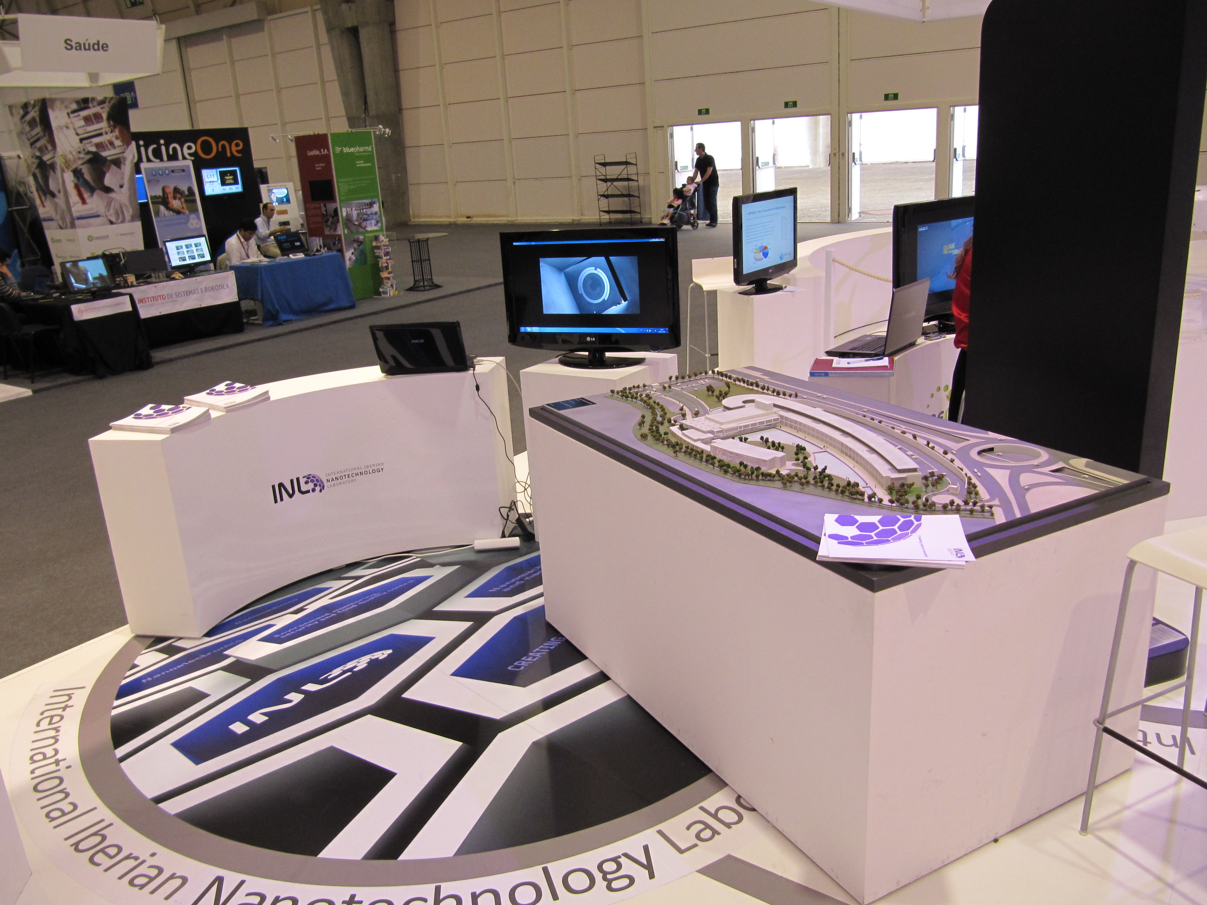
 It’s time to gather again. The International Iberian Nanotechnology Laboratory will hold its second annual meeting, in Braga, (Portugal) next April 12 and 13. The meeting will take place in the new building.
The event will comprise scientific presentations form all INL Post-doc researchers and two Poster Sessions with posters from both INL PhD Programmes.
During the meeting the participants will have the opportunity to make a detailed visit to the INL facilities. INL researchers shall also be updated on the next INL activities. Several members of the INL International Advisory Board, also present in Braga for the annual Advisory Board Meeting, will also attend the meeting, which gives all members of the INL community the opportunity to discuss with them science and their impressions about the INL project.
See you soon in Braga!
INL – News
 New INL researcher Marta Prado
Marta Prado is INL´s latest researcher and has just settled in in Braga. She has an advanced degree in Food Science and Technology and studies in Biology Science from the University of Santiago de Compostela (Spain). Marta has a PhD from the same university in the program of Nutrition, Bromatology and Food technology.
Between the years 1999 and 2006, our new Spanish colleague has been working as a researcher in the Faculty of Veterinary Sciences (Lugo, Spain) from the University of Santiago de Compostela (USC). Between 2006 and 2010, she has been working as Scientific Officer in the Institute of Reference Materials and Measurements from the Joint Research Centre of the European Commission (EC-JRC-IRMM) in Geel, Belgium.
Most of her research experience is related with genomic analysis tools and its application to food analysis, since she had worked on the development and optimization of PCR-based methods for the control of food and animal feeds. In the INL, she will work on the application of magnetic nanobiosensors for the detection of ruminant origin meals in feed.
Recent Comments
MOGAJI Adeola on Marketing of Antimicrobial… updatetoo on Coming Soon: A Foldable i… christopher tingus on Follow the light in the discov… Bangalore Nano on POP – Prototype on … Paul on Solar Energy Backpacks to Rech…
